So if you see tight muscles in one area, you can look at the antagonistic muscles to see why they may actually be over extended and loose, instead of the origin of the problem! You may just be stuck in a compensation pattern. No one is going to want to put their shoulders back if their muscles in their pecs have atrophied and are so tight that it is uncomfortable. So stretching or puffing your chest out may actually be what you need to help the muscles in your shoulders and back! If you have this common issue, and want a cheap fix, laying on a rolled up towel, or a foam roller between your shoulder blades (or scapula) may help this issue correct itself without massage, allowing gravity to do it's work for you.
So let's get into the muscles, and their antagonists. (This will be long, but fear not, you only need to get a general idea of these and their locations, most health professionals will not memorize all of this stuff. . .It is a great resource to keep in your files though. Which is the only reason I have made it!)
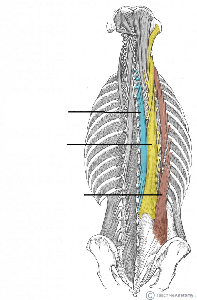 Erector Spinae Group
Action:
- Unilaterally: Laterally flex vertebral column to the same side
- Bilaterally: Extend the vertebral column
Origin: Common tendon that attaches to the posterior surface of sacrum, iliac crest, spinous processes of the lumbar and last two thoracic vertebrae
Insertion: Various attachments at the posterior ribs, spinous and transverse processes of thoracic and cervical vertebrae and mastoid process of temporal bone
Spinalis
Action: Laterally: Flex the head and neck to the same side. Bilaterally: Extend the vertebral column.
Origin:
- Spinous processes of the upper lumbar and lower thoracic vertebrae
- Ligamentum nuchae, spinous process of C-7
Insertion:
- Spinous processes of upper thoracic
- Spinous processes of cervicals, except C-1
Antagonist: Rectus abdominis muscle
Longissiums
Action: Laterally: Flex the head and neck to the same side. Bilaterally: Extend the vertebral column.
Origin: Common tendon, transverse processes of upper five thoracic vertebrae
Insertion:
- Lower nine ribs and transverse processes of thoracic vertebrae
- Transverse processes of cervical vertebrae
- Mastoid process of temporal bone
Antagonist: Rectus abdominis muscle
Iliocostalis
Action: Unilaterally: laterally flex the vertebral column to the same side. Bilaterally: Extend the vertebral column.
Origin: common tendon, posterior surface of ribs 1-12
Insertion:
- Transverse processes of lumbar vertebrae 1-3 and posterior surface of ribs 6-12
- Posterior surface of ribs 1-6
- Transverse processes of lower cervicals
Antagonist: Rectus abdominis muscle
Transversospinalis Group
Multifidi and Rotatores
Action:
- Unilaterally: Rotate the vertebral column to the opposite side
- Bilaterally: Extend the vertebrae
 Origin:
- Multifidi: Sacrum and transverse processes of lumbar through cervical vertebrae
- Rotatores: Transverse processes of lumbar through cervical vertebrae
Insertion: Spinous processes of lumbar vertebrae through second cervical vertebrae
- Multifidi span two to four vertebrae
- Rotatores span one to two vertebrae
 Semispinalis Capitus
Action: Extend the vertebral column and head
Origin: Transverse processes of C-4 to T-5
Insertion: Between the superior and inferior nuchal lines of the occiput
Splenius Capitis and Cervicis
Action:
- Unilaterally: Rotate the head and neck to the same side, laterally flex the head and neck to the same side
- Bilaterally: Extend the head and neck
Origin:
- Capitis: Inferior one-half of ligamentum nuchae and spinous processes of C-7 to T-4
- Cervicis: Spinous processes of T-3 to T-6
Insertion:
- Capitis: Mastoid process and lateral portion of superior nuchal line
- Cervicis: Transverse processes of C-1 to C-3
Antagonist: (Both) Sternocleidomastoid; Longus colli and capitis; Scalenus anterior, medius, and posterior.
(When you think of the area of the neck people would chop to decapitate you, think of the two capitis's)
Quadratus Lumborum

Action:
- Unilaterally:
- Laterally tilt the pelvis
- Laterally flex the vertebral column to the same side
- Assist to extend the vertebral column
- Bilaterally: Fix the last rib during forced inhalation and exhalation
Origin: Posterior iliac crest
Insertion: Last rib and transverse processes of first through fourth lumbar vertebrae
Abdominals
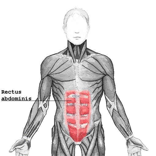
Rectus Abdominis
Action: Flex the vertebral column and tilt pelvis posteriorly
Origin: Pubic crest, pubic symphysis
Insertion: Cartilage of fifth, sixth, and seventh ribs and xiphoid process
Antagonist: Erector spinae, Quadratus Lumborum
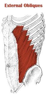
 External Oblique
Action:
- Unilaterally: Laterally flex vertebral column to the same side, Rotate vertebral column to the opposite side
- Bilaterally: Flex the vertebral column, Compress abdominal contents
Origin: External surfaces of fifth to twelth ribs
Insertion: Anterior part of the iliac crest, abdominal aponeurosis to linea alba
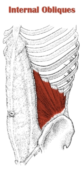
Internal Oblique
Action:
- Unilaterally: Laterally flex vertebral column to the same side, Rotate vertebral column to the same side
- Bilaterally: Flex the vertebral column, Compress abdominal contents
Origin: Lateral inguinal ligament, iliac crest and thoracolumbar fascia
Insertion: Internal surface of lower three ribs, abdominal aponeurosis to linea alba
 Transverse Abdominis
Action: Compress abdominal contents
Origin: Lateral inguinal ligament, iliac crest, thoracolumbar fascia and internal surface of lower six ribs
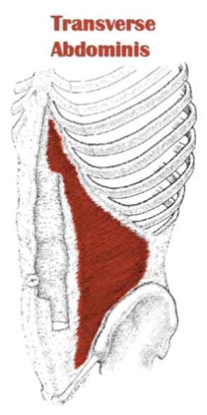 Insertion: Abdominal aponeurosis to linea alba
Antagonist: erector spinae
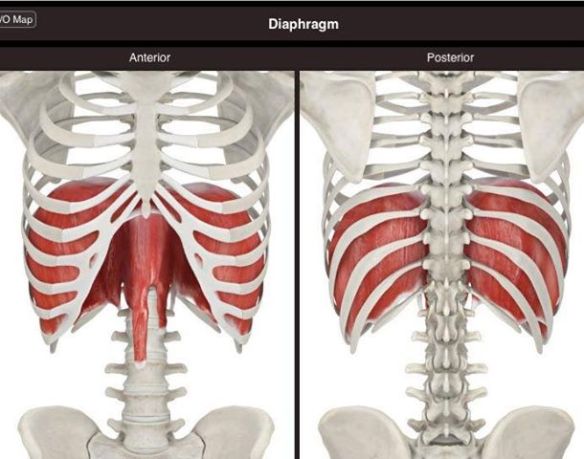
Diaphragm
Action: Draw down the central tendon of the diaphragm, Increase the volume of the thoracic cavity during inhalation
Origin:
- Costal attachment: Inner surface of lower six ribs
- Lumbar attachment: Upper two or three lumbar vertebrae
- Sternal attachment: Inner part of xiphoid process
Insertion: Central tendon
Intercostals
 External and Internal Intercostals
Action:
- External: Draw the ribs superiorly to assist with inhalation
- Internal: Draw the ribs inferiorly to assist with exhalation
Origin: Inferior border of the rib above
Insertion: Superior border of the rib below
Antagonist: External intercostal muscles
Serratus Posterior Superior
 Action: Elevate the ribs during inhalation
Origin: Spinous processes of C-7 to T-3
Insertion: Posterior surface of second through fifth ribs
Serratus Posterior Inferior
Action: Depress the ribs during exhalation
Origin: Spinous processes of T-12 to L-3
Insertion: Posterior surface of ninth through twelfth ribs
Antagonist: Serratus posterior superior, Levatores costarum brevis and longi
 Intertransversarii
Action:
- Unilaterally: Laterally flex the vertebral column to the same side
- Bilaterally: Extend the vertebral column
Origin and Insertion:
- Cervical: Spanning the transverse processes of vertevrae C-2 to C-7
- Lumbar: Spanning the transverse processes of vertebrae L-1 to L-5
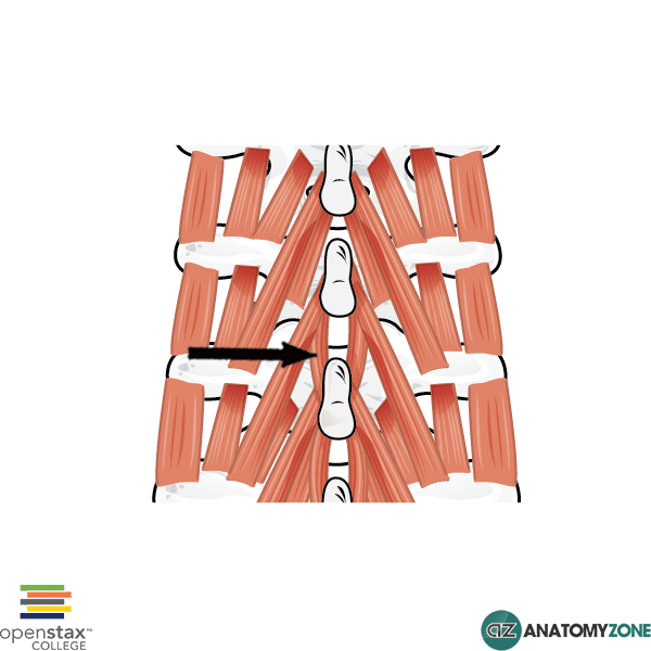 Interspinalis
Action: Extend the vertebral column
Origin and Insertion:
- Cervical: Spanning the spinous processes of C-2 to T-3
- Lumbar: Spanning the spinous processes of T-12 to L-5
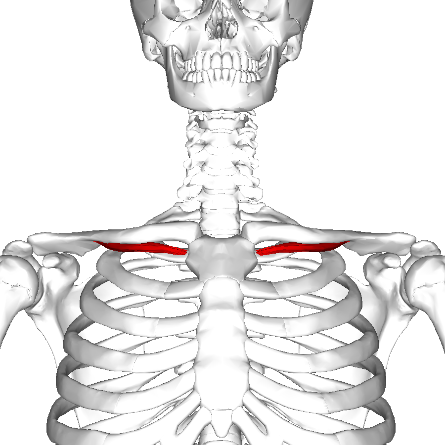 Subclavius (or under the clavicle)
Origin=the first rib right at the junction with the costal cartilage.
Insertion=the groove for the subclavius muscle, called the subclavian groove
Action=depresses the clavicle inferiorly and anteriorly
Antagonist=sternocleidomastoid or SCM muscle
Rhomboid major
origin= spinous process of upper thoracic vertebrae
insertion= vertebral border of scapula
action = rotates and retracts scapula
origin= spinous process of upper thoracic vertebrae
insertion= vertebral border of scapula
action = rotates and retracts scapula
While retracting-Antagonists: Serratus Anterior, Pectoralis Minor
While rotating-Antagonists: Trapezius (upper fibers), Trapezius (lower fibers), Serratus anterior (lower fibers)

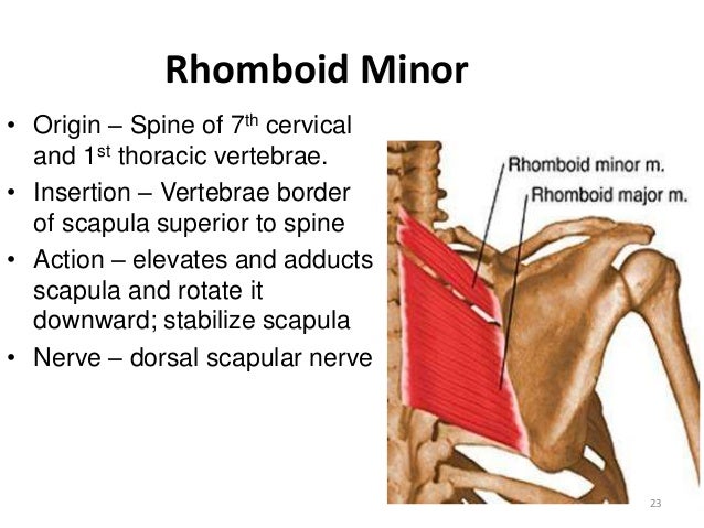
Rhomboid minor
Origin= spinous process of lower cervical and upper thoracic vertebrae
Insertion= vertebral border of scapula action= rotates and retracts scapula
Antagonist= Trapezius

Levetor scapulae
Origin= transverse processes of cervical vertebrae
Insertion= vertebral border of scapula
Action= elevates scapula,
Antagonist=serratus anterior and the lower fibers of the trapezius

Pectoralis minor
Origin= anterior surface of upper-middle ribs


Supraspinatus
Origin= supraspinous fossa of scapula
Insertion= greater tubercle of humerus
Action= abducts arm (part of Rotator cuff)
Antagonist=Pectoralis major lower fibers, latissimus dorsi, teres major


Infraspinatus
Origin= infraspinous fossa of scapula
Insertion= greater tubercle of humerus
Action= laterally rotates arm (part of rotator cuff)
Antagonist=subscapularisteres Major, lats, pec major, anterior deltoid

Insertion= lesser tubercle of humerus
Action= adducts, extends and medially rotates arm
Antagonist=deltoid, infraspinatus, and teres minor.

Teres minor
Origin= On the dorsal surface of the middle half of the lateral border of the scapula.
Insertion=The lowest of the three facets of the greater tubercle of the humerus
Action=Externally rotates the arm
Antagonist= Deltoid

Subscapularis
Origin=Subscapular fossa of the scapula.
Insertion=Lesser tubercle of humerus.
Action=medial rotation of the arm at the shoulder joint
Antagonist= infraspinatus.
While rotating-Antagonists: Trapezius (upper fibers), Trapezius (lower fibers), Serratus anterior (lower fibers)
Rhomboid minor
Origin= spinous process of lower cervical and upper thoracic vertebrae
Insertion= vertebral border of scapula action= rotates and retracts scapula
Antagonist= Trapezius
Levetor scapulae
Origin= transverse processes of cervical vertebrae
Insertion= vertebral border of scapula
Action= elevates scapula,
Antagonist=serratus anterior and the lower fibers of the trapezius
Pectoralis minor
Origin= anterior surface of upper-middle ribs
Insertion= coracoid process of scapula
Action= depresses, protracts, and rotates scapula
Antagonist=rhomboid muscles
Pectoralis major
Origin= clavicle, sternum, and costal cartilages of upper ribs
Insertion= greater tubercle of humerus
Action= flexes, adducts, and medially rotates arm
Action= depresses, protracts, and rotates scapula
Antagonist=rhomboid muscles
Pectoralis major
Origin= clavicle, sternum, and costal cartilages of upper ribs
Insertion= greater tubercle of humerus
Action= flexes, adducts, and medially rotates arm
Antagonist to sterno section= romboid and trapezius muscles
antagonist to the rest are supraspinatus and deltoid muscles
antagonist to the rest are supraspinatus and deltoid muscles

Supraspinatus
Origin= supraspinous fossa of scapula
Insertion= greater tubercle of humerus
Action= abducts arm (part of Rotator cuff)
Antagonist=Pectoralis major lower fibers, latissimus dorsi, teres major
Infraspinatus
Origin= infraspinous fossa of scapula
Insertion= greater tubercle of humerus
Action= laterally rotates arm (part of rotator cuff)
Antagonist=subscapularisteres Major, lats, pec major, anterior deltoid
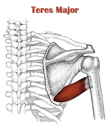
Teres major
Origin= inferior angle of scapula
Origin= inferior angle of scapula
Insertion= lesser tubercle of humerus
Action= adducts, extends and medially rotates arm
Antagonist=deltoid, infraspinatus, and teres minor.

Teres minor
Origin= On the dorsal surface of the middle half of the lateral border of the scapula.
Insertion=The lowest of the three facets of the greater tubercle of the humerus
Action=Externally rotates the arm
Antagonist= Deltoid

Subscapularis
Origin=Subscapular fossa of the scapula.
Insertion=Lesser tubercle of humerus.
Action=medial rotation of the arm at the shoulder joint
Antagonist= infraspinatus.
Trapezius
Action:
 Upper Fibers: elevate and upwardly rotate scapula. Extends neck.
Middle Fibers: Adducts scapula
Lower Fibers: to depress and help upper fibers upwardly rotate scapula
Origin:
Upper part: External occipital proturberance, medial third of the superior nuchal line, the ligamentum nuchae, and the spinous process of C7
Upper part: External occipital proturberance, medial third of the superior nuchal line, the ligamentum nuchae, and the spinous process of C7
Medial Part: Spinous processes of T1 to T5.
Lower Part: Spinous processes of T6 to T12
Insertion:
Upper Part: Lateral third of the clavicle and the medial aspect of the acromion process of the scapula
Upper Part: Lateral third of the clavicle and the medial aspect of the acromion process of the scapula
Middle Part: Medial edge of the superior surface of the acromion process of the scapula and the superior edge of the scapular spine.
Lower Part: Tubercles of the apex of the scapular spine.
And that's all folks! (For now at least. . .)