Friday, September 11, 2015

Leg and foot muscles: origins, insertions and actions.


Just in case you are looking for it all in one place, and easily copy and pastable (like I was. . .and did not find) check out the Leg and foot muscles, and their origins, insertions and actions. And to start us off:

Gastocnemius

  Action:  Flex the knee, plantar flex the ankle

  Origin:  Condyles of the femur, posterior surfaces

  Insertion:  Calcaneus via calcaneal tendon

Soleus

  Action:  Plantar flex the ankle

  Origin:  Soleal line; proximal, posterior surface of tibia and posterior aspect of head of fibula

  Insertion:  Calcaneus bia calcaneal tendon

Plantaris

  Action:  Weak plantar flexion of the ankle, weak flexion of the knee

  Origin:  Lateral supracondylar line of femur

  Insertion:  Calcaneus via calcaneal tendon


Popliteus

  Action:  Medially rotate the flexed knee, flex the knee

  Origin:  Lateral condyle of the femur

  Insertion:  Proximal, posterior aspect of tibia


Peroneus Longus

  Action:  Evert the foot, assist to plantar flex the ankle

  Origin:  Head of fibula and proximal two-thirds of lateral fibula

  Insertion:  Base of the first metatarsal and medial cuneiform

Peroneus Brevis

  Action:  Evert the foot, assist to plantar flex the ankle

  Origin:  Distal two-thirds of lateral fibula

  Insertion:  Tuberosity of fifth metatarsal

Extensors of the Ankle and Toes

Tibialis Anterior

  Action:  Invert the foot, Dorsiflex the ankle

  Origin:  Lateral condyle of tibia; proximal, lateral surface of tibia and interosseous membrane

  Insertion:  Medial cuneiform and base of the first metatarsal

Extensor Digitorum Longus

                                                                 Action:
  • Extend the second through fifth toes
  • Dorsiflex the ankle
  • Evert the foot
  Origin:  Lateral condyle of tibia; proximal, anterior shaft of fibula and interosseous membrane

  Insertion:  Middle and distal phalanges of second through fifth toes


Extensor Hallucis Longus

  Action:  
  • Extend the first toe
  • Dorsiflex the ankle
  • Invert the foot
  Origin:  Middle, anterior surface of fibula and interosseous membrane

  Insertion:  Distal phalanx of first toe

Flexors of the Ankle and Toes


Tibialis posterior

Action: Invert foot and plantar flex ankle

Origin: Proximal post. shaft of tibia, proximal fibula and interosseous membrane

Insertion: Navicular cuneiforms, cuboid and bases of second through fourth metatarsals


Flexor digitorium longus

Action: Flex second through fifth toes, invert foot and weak plantar flexion of ankle

Origin: Middle post. surface of tibia

Insertion: Distal phalanges of second through fifth toes


Flexor Hallucis Longus

Action: Flexfirst toe, invert foot and weak plantar flexion of ankle

Origin: Middle half of post. fibula

Insertion: Distal phalanges of first toe

Extensor digitorum longus

Action: Extends toes and extends foot at ankle
Origin:Upper two thirds of anterior shaft of fibula, interosseous membrane and superior tibiofibular joint
Insertion: Extensor expansion of lateral four toes


Extensor Digitorum Brevis

Action: Extend second through fourth toes

Origin: Calcaneus

Insertion: Second through fourth toes via extensor digitorum longus tendons


Flexor Digitorum Brevis

Action: Flex middle phalanges of second through fifth toes

Origin: Calcaneus

Insertion: Middle phalanges of second through fifth toes


Abductor Hallucis

Action: Abduct first toe and assist to flex first toe

Origin: Calcaneus

Insertion: Proximal phalange of first toe


Abductor Digiti Minimi

Action: Flex fifth toe and assist to abduct fifth toe

Origin: Calcaneus 

Insertion: Proximal phalange of fifth toe


Extensor Hallucis Brevis

Action: Extend first toe

Origin: Calcaneus

Insertion: Proximal phalange of first toe


Flexor Hallucis Brevis

Action: Flex first toe

Origin: Plantar surfaces of cuboid and lateral cuneiform

Insertion: Med. and lat. sides of base of proximal phalange of first toe

Adductor Hallucis

Action: Adduct first toe and assist to maintain transverse arch of foot

Origin: Oblique head- Bases of second through fourth metatarsals
Transverse head- Platar ligament of third through fifth metotarsophalangeal joints

Insertion: Lat. surface of base of proximal phalange os first toe and lateral sesamoid bone


Abductor Hallucis
Action: Flexes and abducts big toe. Supports medial longitudinal arch

Origin: Medial process of posterior calcaneal tuberosity & flexor retinaculum

Insertion: Medial aspect of base of proximal phalanx of big toe via medial sesamoid


Flexor digiti minimi brevis

Action: Flexes metatarsophalangeal joint of little toe

Origin: Base of 5th metatarsal and sheath of peroneus longus

Insertion: Lateral side of base of proximal phalanx of little toe


Quadratus Plantae

Action: Assist flexor digitorum longus to flex second through fifth toes

Origin: Platar surface of calcaneus

Insertion: Post., lat. aspect of flexor digitorum longus tendon


Lumbricals 

Action: Flex proximal phalanges of second through fifth toes at metatarsophalangeal joints and extend middle and distal phalanges of second through fifth toes and interphalangeal joints

Origin: Tendons of flexor digitorum longus

Insertion: Bases of proximal Phalanges of second through fifth toes and expansions of extensor digitorum longus tendons


Plantar Interossei

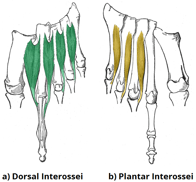
Action: Adduct and flex third through fifth toes

Origin: Bases of third through fifth metatarsals

Insertion: Medial surfaces of proximal phalanges of third through fifth toes


Dorsal Interossei

Action: Abduct and flex second through fourth toes

Origin: Shafts of first through fourth metatarsals

Insertion: First- Medial surface of proximal phalange of second toe
Second through fourth- Lateral surfaces of proximal phalange of second through fourth toes

I hope this was a help to you as just making it has been to me. Please comment below if I missed anything or said something wrong, as I simply am copying others work here, and don't know this stuff myself yet.

Muscles of the Pelvis and Thigh


Muscles of the Pelvis & Thigh.


Tensor Fasciae Latae

Action: Flex, medially rotate, and abduct the hip

Origin: Iliac crest, posterior to the ASIS

Insertion: Iliotibial tract


Iliotibial Tract

Superficial sheet of fascia (not technically a muscle) that covers the vastus lateralis – emerges from the Gluteal fascia.

Sartorius

Action: 
  • Flex, laterally rotate, and abduct the hip
  • Flex the knee
  • Medially rotate the flexed knee
Origin: Anterior superior iliac spine

Insertion: Proximal, medial shaft of the tibia at pes anserinus tendon

Quadriceps group (4)
Action:  

  • Extend the knee (tibiofemoral joint) -- all
  • Flex the hip -- Rectus Femoris
Origin:
  • Rectus Femoris: Anterior inferior iliac spine (AIIS)
  • Vastus Medialis: Medial lip of linea aspera
  • Vastus Lateralis: Lateral lip of linea aspera, gluteal tuberosity and greater trochanter
  • Vastus Intermedius: Anterior and lateral shaft of the femur
Insertion: Tibial tuberosity (via the patella and patellar ligament)

Hamstring group (3)

Biceps Femoris

Action:
  • Flex the knee
  • Laterally rotate the flexed knee
  • Long head: Extend the hip and assist to laterally rotate the hip
  • Tilt the pelvis posteriorly
Origin:
  • Long head: Ischial tuberosity
  • Short head: Lateral lip of linea aspera

Insertion: Head of the fibula


Semitendinosus

Action:
  • Flex the knee
  • Medially rotate the flexed knee
  • Extend the hip
  • Assist to medially rotate the hip
  • Tilt the pelvis posteriorly
Origin: Ischial tuberosity


Insertion: Proximal, medial shaft of the tibia at pes anserinus tendon


Semimembranosus

Action:

  • Flex the knee
  • Medially rotate the flexed knee
  • Extend the hip
  • Assist to medially rotate the hip
  • Tilt the pelvis posteriorly
Origin: Ischial turberosity

Insertion: Posterior aspect of medial condyle of tibia


Gluteals
Gluteus Maximus

Action:
  • Extend the hip
  • Laterally rotate the hip
  • Abduct the hip
  • Adduct the hip (lower fibers)
Origin:
  • coccyx
  • edge of sacrum
  • posterior iliac crest
  • sacrotuberous and sacroiliac ligaments

Insertion: Iliotibial tract (upper fibers) and gluteal tuberosity (lower fibers)


Gluteus Medius

Action:  
  • All fibers: Abduct the hip
  • Anterior fibers: Flex and medially rotate the hip
  • Posterior fibers: Extend and laterally rotate the hip
Origin:  
  • Gluteal surface of ilium
  • between posterior and anterior gluteal lines
  • just below the iliac crest
Insertion: Lateral aspect of greater trochanter


Gluteus Minimus

Action: Abduct, medially rotate, and flex the hip


Origin: Gluteal surface of the ilium between the anterior and inferior gluteal lines

Insertion: Anterior aspect of greater trochanter


Adductor Group

Actions:

All: Adduct and medially rotate the hip

All except Gracilis: Assist to flex the hip


      Gracilis: Flex and medially rotate the flexed knee


   Posterior fibers of Adductor Magnus: Extend the hip


Adductor Magnus

Origin: Inferior ramus of the pubis, ramus of ischium and ischial tuberosity

Insertion: Medial lip of linea aspera and adductor tubercle


Adductor Longus

Origin: Pubic tubercle

Insertion: Medial lip of linea aspera


Adductor Brevis

Origin: Inferior ramus of pubis

Insertion: Pectineal line and medial lip of linea aspera


Pectineus

Origin: Superior ramus of pubis

Insertion: Pectineal line of femur


Gracilis

Origin: Inferior ramus of pubis

Insertion: Proximal, medial shaft of tibia at pes anserinus tendon


Lateral Rotators of the Hip

Action:  

  • All laterally rotate the hip at the coxal joint
  • Piriformis abducts the hip when the hip is flexed.


Piriformis

 Origin: Anterior surface of sacrum

Insertion: Superior aspect of greater trochanter


Quadratus Femoris

Origin: Lateral border of ischial tuberosity

Insertion: Intertrochanteric crest, between the greater and lesser trochanters


Obturator Internus

Origin: Obturator membrane and inferior surface of obturator foramen

Insertion: Medial surface of greater trochanter


Obturator Externus

Origin: Rami of pubis and ischium, obturator membrane

Insertion: Trochanteric fossa of femur


Gemellus Superior

Origin: Ischial spine

Insertion: Medial surface of greater trochanter


Gemellus Inferior

Origin: Ischial tuberosity

Insertion: Medial surface of greater trochanter

Iliopsoas group

Psoas minor

Action: Posterior rotation of pelvis.

Origin: Anterolateral surface of T12 and L1 vertebrae.

Insertion: Superior ramus of pubis. Action: Posterior rotation of pelvis.

Psoas Major

Action:
  • With the origin fixed: flex the hip, may laterally rotate the hip
  • With the insertion fixed: flex the trunk toward the thigh, tilt pelvis anteriorly
  • Unilaterally: Assist to laterally flex the lumbar spine
Origin: Bodies and transverse processes of lumbar vertebrae

Insertion: Lesser trochanter


Iliacus

Action:

  • With the origin fixes: flex the hip, may laterally rotate the hip
  • With the insertion fixed: flex the trunk toward the thigh, tilt pelvis anteriorly
Origin: Iliac fossa

Insertion: Lesser trochanter

That's all of them!

Thursday, September 10, 2015

Head, neck and face muscles: action, origin and insertion.


Muscles – Head, Neck, & Face

Sternocleidomastoid


Action: 

Unilateral
  • laterally flex the head and neck to the same side
  • rotate the head and neck to the opposite side
Bilateral
  • flex the neck
  • assist to elevate the rib cage during inhalation

Origin:  

  • Sternal Head: top of manubrium
  • Clavicular Head: medial 1/3 of the clavical

Insertion:


Mastoid process of temporal bone and the lateral portion of superior nuchal line of occiput


Scalenes

Action: Same for all the scalenes.

Unilateral
  • With the rib fixed, laterally flex the head and neck to the same side.
  • Rotate head and neck to the opposite side.

Bilateral
  • Elevate the ribs during inhalation.
  • Flex the head and neck (anterior).


Anterior Scalene

Origin: Transverse processes of third through sixth cervical vertebrae.

Insertion: First rib.


Middle Scalene

Origin: Transverse processes of second through seventh cervical vertebrae.

Insertion: First rib.


Posterior Scalene

Origin: Transverse processes of sixth and seventh cervical vertebrae.

Insertion: Second rib.


Masseter

Action: Elevate the mandible. May assist to protract the mandible.

Origin: Zygomatic arch.

Insertion: Angle and ramus of mandible.


Temporalis

Action: Elevates and retracts the mandible.

Origin: Temporal fossa and fascia.

Insertion: Coronoid process and anterior edge of ramus of the mandible.

Digastric

Action:  
  • With hyoid bone fixed, depresses the mandible.
  • With mandible fixed, elevates the hyoid bone.
  • Retracts the mandible.

Origin: Mastoid process.

Insertion: Inferior border of the mandible.

Platysma

Action:
  • Assist to depress the mandible.
  • Tighten the fascia of the neck.
  • Draw down the corner of the mouth.

Origin: Fascia covering superior part of pectoralis major.

Insertion: Base of mandible, skin of lower part of face.

Occipitofrontalis

Action:  
  • Frontalis: Raise the eyebrows and wrinkle the forehead.
  • Occipitalis: Anchor and retract the galea posteriorly.

Origin: Both: Galea apponeurotica

Insertion:  
  • Frontalis: Skin superior to eyebrows.
  • Occipitalis: Superior nuchal line of the occiput

Medial Pterygoid

Action:  
  • Unilateral: Laterally deviates the mandible to the opposite side.
  • Bilaterally: Elevates and protracts the mandible.

Origin: Medial surface of lateral pterygoid plate of sphenoid and tuberosity of maxilla.

Insertion: Medial surface of ramus of the mandible.

Lateral Pterygoid

Action:  
  • Unilateral: Laterally deviates the mandible to the opposite side.
  • Bilateral: Protracts the mandible.
Origin:  
  • Superior head: Infratemporal surface and crest of greater wing of sphenoid bone.
  • Inferior head: Lateral surface of lateral pterygoid plate of sphenoid bone.
Insertion: Articular disk and capsule of temporomandibular joint, neck of mandible.

Longus Capitis

Action:  
  • Unilateral: Laterally flexes the head and neck to the same side.
  • Bilaterally: Rotates the head and neck to the same side.
Origin: Transverse processes of C-3 through C-6.
Insertion: Inferior surface of occiput.

Longus Colli

Action:
  • Unilateral: Laterally flexes the head and neck to the same side.
  • Bilaterally: Rotates the head and neck to the same side.
Anterior View of Rectus Capitis Anterior
Origin: Bodies of C-5 through T-3, transverse processes of C-3 through C-5.

Insertion:  
  • Tubericle on anterior arch of the atlas
  • Bodies of the axis, C-3 & C-4
  • Transverse processes of C-5 & C-6
Rectus capitis anterior

Action: Aids in flexion of the head and the neck
Origin: Anterior surface of the lateral mass of the atlas (C1 vertebra) and the root of its transverse process[
Insertion: The inferior surface of the occipital bone anterior to the foreamen magnum

Some of these muscles are grouped into muscle groups,
below are some.

Infrahyoids (inferior to hyoid)

Action: Depress the hyoid bone and thyroid cartilage.

Origin:  
  • Sternohyoid & Sternothyroid: Top of manubrium.
  • Thyrohyoid: Thyroid cartilage.
  • Omohyoid: Superior border of the scapula.

Insertion:  
  • Sternothyroid: Thyroid cartilage.
  • Sternohyoid, Thyrohyoid, & Omohyoid: Hyoid bone.
Suprahyoids (superior to hyoid)

Action: Elevate hyoid and tongue. Depresses mandible.

Origin:  
  • Geniohyoid & Mylohyoid: Underside of mandible.
  • Stylohyoid: Styloid process.
Insertion: Hyoid bone.

Suboccipitalis (under the occipital bone) group of 4

Rectus Capitis Posterior Major
Action: Extends and rotates atlanto-occipital joint
Origin: Spinous process of axis (C2)
Insertion: Lateral half of inferior nuchal line
        Antagonist: Extension – Longus Capitis, Rectus Capitis Anterior; Rotation – Oblique Capitis Inferior, Rectus Capitis Posterior Major; Lateral flexion – Oblique Capitis Superior, Rectus Capitis Lateralis.

Rectus Capitis Posterior Minor
Action: Extends and rotates atlanto-occipital joint
Origin: Posterior process of atlas (C1)
Insertion: Medial half of inferior nuchal line
        Antagonist: 

Oblique Capitis Superior
Action:
Origin:
Insertion:
       Antagonist:

Oblique Capitis Inferior
Action:
Origin:
Insertion:
       Antagonist:


And that is more than you will likely ever need to know about the head, neck and face.